Definition
An ulnar fracture occurs when the ulna, one of the two bones in the forearm, breaks. The ulna is located on the side of the forearm closest to the little finger, opposite the radius, which is located near the thumb. The fracture can occur in any part of the ulna, including near the wrist, in the middle, or near the elbow.
Ulnar fractures are frequently seen in combination with radius fractures, although isolated ulna fractures are less common.
Causes
Ulna fractures are typically caused by either direct or indirect trauma. Direct traumas occur when the forearm is struck by an object, such as being hit by a ball during sports, struck by a blunt force, or involved in a car accident.
Meanwhile, indirect traumas are often caused by falls, especially those from a height where the person lands on an outstretched arm.
Risk Factor
The risk of an ulnar fracture is higher in active adolescents, particularly those who play sports and are prone to both direct and indirect forearm injuries. Adults with osteoporosis are also more susceptible to these fractures due to weakened bones.
Symptoms
The signs of an ulnar fracture typically include:
-
Pain: The pain often intensifies with movement of the hand or forearm.
-
Swelling: The forearm may become swollen and firm to the touch.
-
Deformity: There may be an abnormal angle at the wrist or a visible bone protrusion.
-
Open fracture: In some cases, the broken bone may pierce the skin, creating an open wound at the fracture site.
-
Loss of function: The fracture can result in limited movement in the hand and forearm.
Diagnosis
A doctor typically diagnoses an ulnar fracture through a combination of medical history, physical examination, and additional tests on the forearm.
Medical Interview
During the interview, the doctor will ask questions to gather important information, including:
-
Current symptoms and their severity
-
Any prior injuries or similar incidents
-
Occupational and daily activities that might affect the injury
-
Past medical history, including any relevant conditions
-
Family history of bone or joint issues
Physical Examination
After the interview, the doctor will conduct a general physical examination to check vital signs such as blood pressure, respiratory rate, pulse rate, and body temperature. The doctor will also carry out a specific examination focused on the injured area.
A physical exam of the forearm in someone with an ulnar fracture may reveal signs of deformity, discoloration of the skin, or open wounds. The doctor will also check for symptoms of compartment syndrome, a serious condition where pressure builds within the muscles of the forearm, possibly impairing circulation.
Additionally, the doctor will assess nerve and blood vessel function by checking pulses at the thumb and little finger and testing sensory and motor functions to evaluate nerve function. This helps ensure that the blood vessels of the radius and ulna are not compromised and that there is no nerve damage due to the fracture.
Diagnostic Tests
To confirm an ulnar fracture, your doctor may order diagnostic tests, such as X-rays or CT scans, to visualize the forearm, rule out other injuries, and ensure the correct diagnosis. Standard X-ray views typically include front, side, and oblique images of the forearm to assess the location and extent of the fracture.
Management
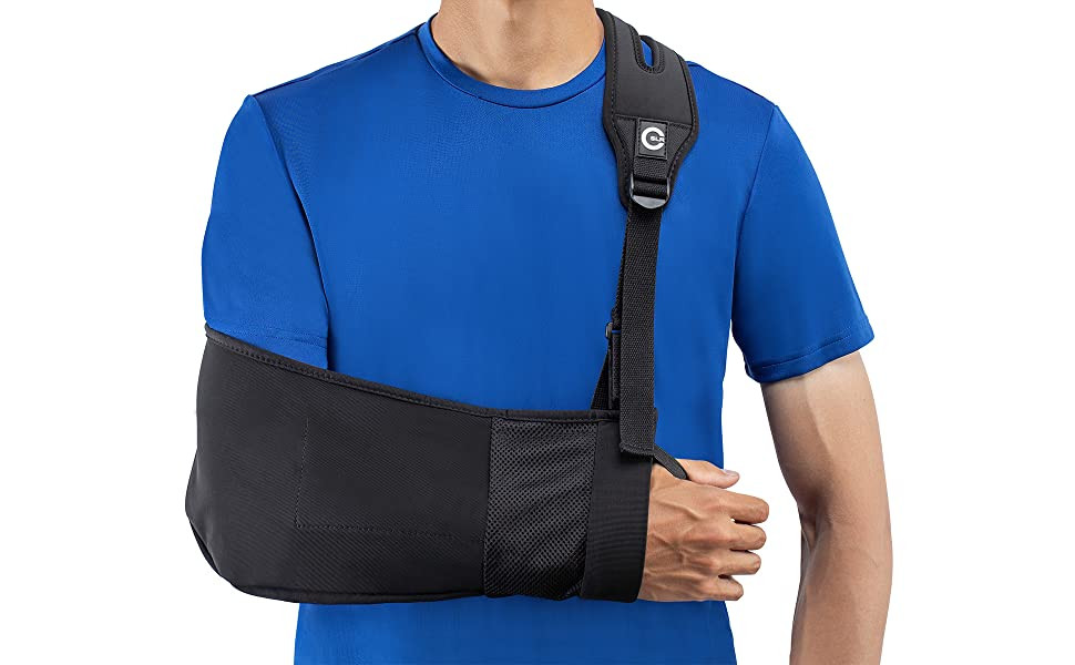
The treatment for an ulnar fracture depends on the severity and type of fracture. It can be either non-surgical or surgical. In some cases, non-surgical treatment may be enough to fully heal the fracture, particularly in children with simpler ulnar fractures or in adults with stable fractures of the ulna without involving the radius.
Non-Surgical Treatments
Non-surgical treatments usually involves wearing a functional cast or brace for about 4 to 6 weeks to immobilize the forearm. The patient is advised to avoid bearing weight on the injured arm during this period. The doctor will monitor the healing progress through periodic X-rays and physical exams. Once the cast is removed, physical therapy, along with a gradual return to weight-bearing activities, is recommended to restore full range of motion in the forearm.
Surgical Treatments
In cases where non-surgical treatment is not sufficient, such as for adults with complex fractures or open fractures, surgery is generally required. The surgical technique will depend on the nature of the fracture and the affected bone. Some of the procedures include:
-
Open Reduction and Internal Fixation: This involves making an incision to access the broken bone, realigning it, and securing it with screws or plates.
-
External Fixation: A plate and screws are attached to the outside of the forearm to stabilize the bone from the outside.
-
Intramedullary Nailing: This technique involves inserting a metal rod through the center of the bone to provide internal support.
Surgical treatment for open fractures often includes bone alignment, cleaning of any wounds, and the use of antibiotics to prevent infection.
After surgery, the forearm will typically be immobilized in a splint or cast to allow the bone to heal. Weight-bearing should be avoided for approximately six weeks. After this period, range-of-motion exercises will begin under the supervision of a doctor, with regular follow-up appointments to monitor the healing process through physical exams and radiological tests.
Healing from an ulnar fracture typically takes 2 to 3 months, but the exact healing time can vary depending on the complexity of the fracture and individual factors such as medical history and adherence to treatment.
Complications
While many ulnar fractures heal without issues, several complications can arise during the healing process, including:
-
Infection: This is more common in open fractures, especially in procedures like open reduction and internal fixation. The incidence of infection in these surgeries is about 3%.
-
Compartment Syndrome: This serious condition, where pressure builds up in the muscle compartments of the forearm, can occur in up to 15% of cases, depending on the fracture's nature. Factors like high-energy injuries, open fractures, blood vessel injuries, and blood clotting disorders increase the risk of compartment syndrome.
-
Nonunion: When a fracture fails to heal, it is referred to as nonunion. This risk is higher in comminuted fractures (where the bone is broken into multiple pieces) or when the placement of internal fixation plates is improper.
-
Malunion: This occurs when the broken bone heals in an incorrect position, leading to misalignment.
-
Nerve and Blood Vessel Injury: The fracture or surgical procedure may cause damage to the nearby nerves and blood vessels, which can affect the function of the forearm and hand.
-
Refracture: Refracture happens when the bone breaks again before it has fully healed. Premature removal of plates or large plates used in the fixation process can increase the risk of refracture. Studies show that about 10% of cases with premature plate removal result in refracture.
Prevention
While it is difficult to completely prevent fractures due to the sudden nature of accidents, there are several steps you can take to reduce your chances of experiencing a fracture:
-
Calcium and Vitamin D: Ensure you're consuming enough calcium and vitamin D, as these nutrients are essential for bone strength and health. Follow your doctor’s recommendations for the appropriate intake of these nutrients.
-
Protective Gear: Use appropriate protective equipment, such as wrist and forearm guards, during sports to protect your bones from impact.
-
Weight-Bearing Activities: Engage in weight-bearing physical exercises, which help strengthen the muscles around bones and joints, providing additional support and protection.
Postmenopausal women and men aged 70 or older should consult with a doctor for an osteoporosis evaluation. If you are at risk for osteoporosis, your doctor may recommend a screening test. Early detection and treatment can prevent fractures and other related complications.
When to See a Doctor?
Although an ulnar fracture is generally not life-threatening, certain situations can lead to rapid infection, which can result in serious complications. Seek immediate medical help if you or someone you know experiences any of the following symptoms:
-
A bone breaking through the skin in the forearm or wrist
-
A deep wound with exposed skin on the forearm or wrist, potentially indicating a fracture
Looking for more information about other diseases? Click here!
- dr Hanifa Rahma
Distal ulna fracture: What is it, how is it managed, and more - osmosis (no date). Available at: https://www.osmosis.org/answers/distal-ulna-fracture (Accessed: November 28, 2022).
Isolated ulnar shaft fracture (no date) Orthobullets. Available at: https://www.orthobullets.com/trauma/422969/isolated-ulnar-shaft-fracture (Accessed: November 28, 2022).
Sarah Lewis, P.D. (2020) Forearm fracture, Healthgrades. Healthgrades. Available at: https://www.healthgrades.com/right-care/bones-joints-and-muscles/forearm-fracture (Accessed: November 28, 2022).

/62d2449c2c10c.jpg)